Single-shot hyperspectral wavefront imaging
Blochet, B., N. Lebas, P. Berto, D. Papadopoulos, and M. Guillon
Nature Communications 17, no. 1 (2026)

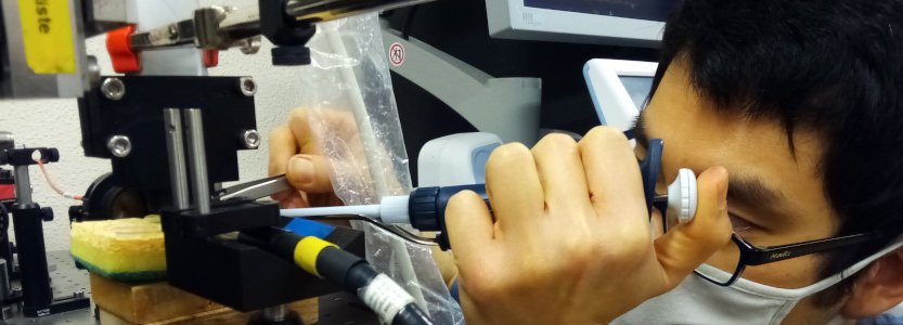
Résumé: Single-shot hyperspectral wavefront sensing is essential for applications like spatio-spectral coupling metrology in high-power laser or fast material dispersion imaging. Under broadband illumination, traditional wavefront sensors assume an achromatic wavefront, which makes them unsuitable. We introduce a hyperspectral wavefront sensing scheme based on the Hartmann wavefront sensing principles, employing a multicore fiber as a Hartmann mask to overcome these limitations. Our system leverages the angular memory effect and limited spectral correlation width of the multicore fiber, encoding wavefront gradients into displacements and the spectral information into uncorrelated speckle patterns. This method retains the simplicity, compactness, and single-shot capability of conventional wavefront sensors, with only a slight increase in computational complexity. It also allows a tunable trade-off between spatial and spectral resolution. We demonstrate its efficacy for recording the hyperspectral wavefront cube from single-pulse acquisitions at the Apollon multi-petawatt laser facility, and for performing multispectral microscopic imaging of dispersive phase objects.
|


|
Time-reversal lens and phase conjugation with an active spatiotemporal interface [Invited]
Wildeman, S., A. Eddi, and E. Fort
Optical Materials Express 16, no. 10, 3231-3239 (2026)
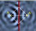
Résumé: Active spatiotemporal interfaces enable wave control through frequency conversion, phase conjugation, and negative refraction. Thin time-modulated interfaces have been predicted to act as flat time-reversal lenses, forming phase-conjugated images on the far side of the interface, yet experiments have mostly addressed phase conjugation or negative refraction in simple beam geometries rather than imaging. Here we realize a flat time-reversal lens for water waves using a subwavelength electrostrictive interface that induces up to a twofold local change in wave velocity. We demonstrate diffraction-limited image formation with a focal spot of half the source wavelength, tunable imaging at complementary frequencies, and control of object-image relations using curved interfaces.
|
|
A floating body with no preferred orientation: An experimental realization
Pontiggia, L., A. Campaniello, and E. Fort
American Journal of Physics 94, no. 10, 853-858 (2026)
Résumé: Editor's Note: Few students will be surprised to hear that a buoyant sphere can float in any orientation. What might astonish them is the fact that there are other shapes that can float with no preferred orientation! Teachers covering buoyancy will appreciate this striking demonstration and simple tabletop experiment: a heart-shaped object that can settle at any angle.
|
|
In-plane vector-field imaging of propagating surface phonon polaritons [Invited]
Niemann, R., D. S. Mader, G. Álvarez-Pérez, A. I. F. Tresguerres-Mata, A. Tarazaga Martín-Luengo, S. Partel, J. D. Caldwell, N. S. Mueller, M. Wolf, J. Martín-Sánchez, P. Alonso-González, and A. Paarmann
Optical Materials Express 16, no. 10, 3483-3495 (2026)
Résumé: Polariton interferometry through optical near-field microscopy has become a powerful tool in nanophotonics, enabling direct spatial access to the propagation characteristics of strongly confined, evanescent polariton modes. Scattering-type near-field optical microscopy has matured as the prime tool for such studies, yet mostly the out-of-plane components of the optical near fields are probed, owing to the elongated geometry of the nanotip. Here, we demonstrate a complementary far-field nonlinear microscopy approach that allows selective probing of in-plane polariton field components. Accessing the full vector field is interesting when studying complex mode patterns such as hyperbolic polaritons or skyrmions, where the in-plane field components are typically only inferred from the out-of-plane component but not measured directly. To this end, we use nonlinear infrared-visible wide-field sum-frequency generation microscopy, where the short visible wavelength of the nonlinear signal provides the high spatial resolution to access evanescent modes in the infrared. The symmetry selection rules of the nonlinear process further enable polarization-selective imaging of both in-plane polariton field components through spatial interferometry. The concept is demonstrated experimentally using surface phonon polaritons at the AlN-air interface launched by a gold antenna. A simple, semi-analytical model reproduces the peculiar propagation patterns. Hyperspectral imaging with a tunable narrowband laser further gives access to the polariton dispersion. The wide-field methodology holds high promise for in-depth and high-throughput studies of infrared nanophotonic structures.
|
|
Incidental Observations of Acoustic Droplet Vaporization-Induced Hemorrhage Using PFH Droplets
Teston, E., A. Aissani, S. Décombas-Deschamps, V. Hingot, F. Lager, G. Renault, and O. Couture
Ultrasound in Medicine and Biology 52, no. 9, 2133-2139 (2026)
|
|
The Organization of Choroidal Arteries in Normal Eyes
Barre, A., C. Legall, C. Chaumette, D. C. Farias, S. Mrejen, L. Puyo, M. Atlan, and M. Paques
Ophthalmology Science 6, no. 9 (2026)
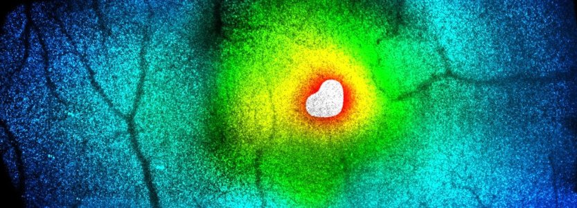
Résumé: Objective: To determine the organization of normal human choroidal arteries using a combination of laser Doppler holography (LDH) and OCT. Design: Cross-sectional clinical study. Subjects: One hundred thirty-two eyes of 74 healthy subjects. Methods: By combining images from a prototypic LDH system and from OCT, we documented the 3-dimensional disposition of choroidal arteries from their emergence from ciliary arteries to precapillary arterioles. Main Outcome Measures: Distribution, diameters, and pathways of choroidal arteries. Results: A notable intereye variability in the disposition of arteries was found, in which dominant patterns were identified. A short ciliary artery emerging within 1000 μm from the fovea was identified in 63% of eyes, giving rise to a horizontal submacular artery (SMA; mean diameter 119.7 ± 24.9 μm) oriented temporally. The detection rate of an SMA was inversely correlated with choroidal thickness, that is, SMAs were more often identified in thinner choroids. Among cases with a detectable SMA, its diameter positively correlated with choroidal thickness (P < 0.003). Long posterior ciliary arteries were identified in 43% of eyes and emerged at a mean distance of 4820 ± 1567 μm temporal to the fovea (mean diameter 115 ± 41 μm). Paraoptic arteries radiated from the margins of the optic nerve head, some of which perfused the macula. Disseminated axially oriented arterioles perfusing precapillary arterioles were detected from first-order arteries. Conclusions: Using a combination of LDH and OCT in healthy eyes, we identified recurrent organizational patterns of choroidal arteries in normal eyes, including SMAs, long posterior ciliary arteries, paraoptic arteries, and precapillary complexes. Characterizing the physiologic variants of the disposition of choroidal arteries helps to provide a reference framework that will enable the identification of disease-related changes. Financial Disclosure(s): Proprietary or commercial disclosure may be found in the Footnotes and Disclosures at the end of this article.
|
|